🏆 Medical Achievement
Surgery
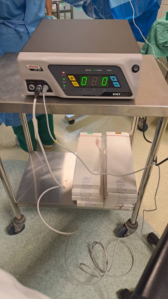
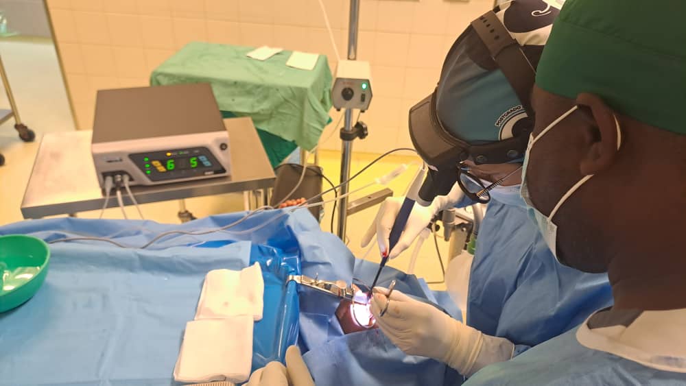
First tonsillectomy by radiofrequency in Equatorial Guinea
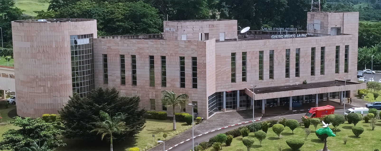
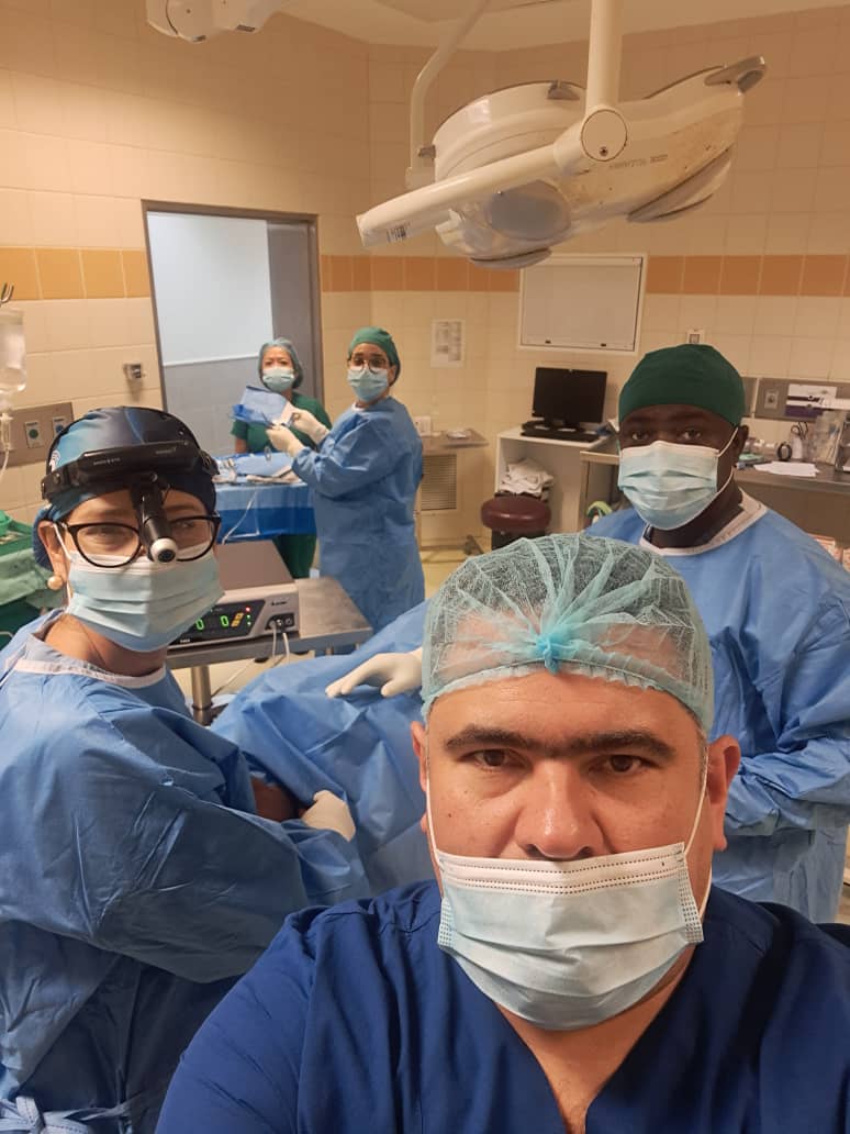
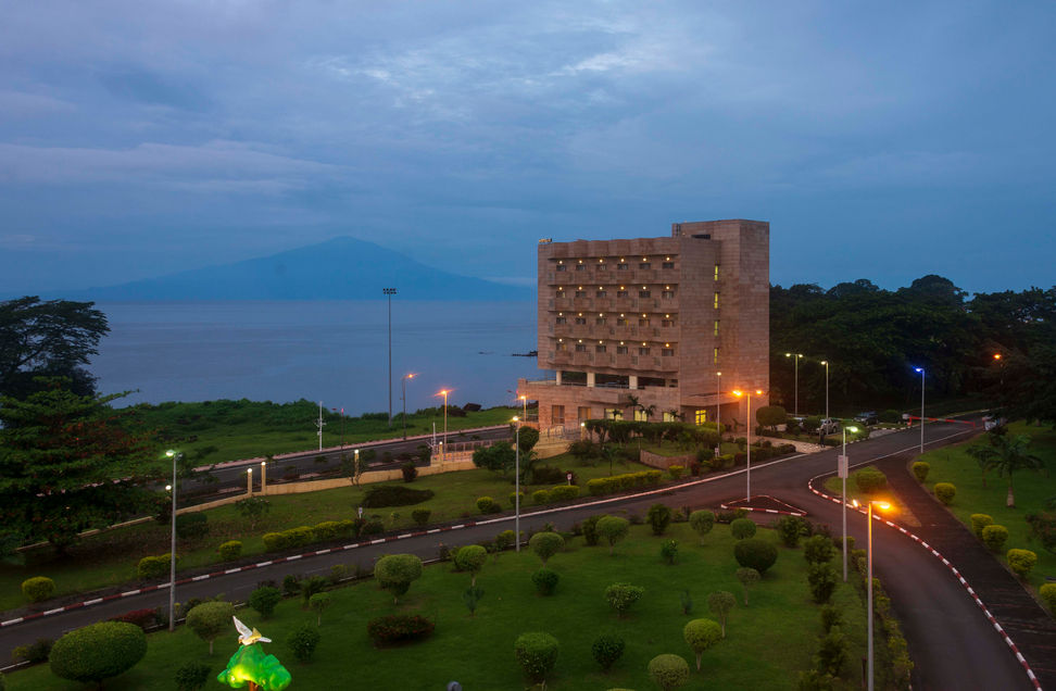
Today, November 13, 2025, the first radiofrequency tonsillectomy in Equatorial Guinea was successfully performed at La Paz Medical Center in Malabo. Surgeon: Dr. Rosalinda Peruzini. Assistant: Dr. Juan Carlos Abeso. Anesthesiologist: Dr. Hector Rojas.